Voluntary control of skeletal muscles allows humans to perform intentional movements such as walking writing and lifting objects. Unlike involuntary muscles which function automatically skeletal muscles are consciously controlled by the nervous system.
The primary system responsible for voluntary muscle control is the somatic nervous system (SNS). This system transmits signals from the brain and spinal cord to skeletal muscles allowing precise movements. In this topic we will explore how voluntary muscle control works the role of different brain regions and the importance of neurons in movement coordination.
What Are Skeletal Muscles?
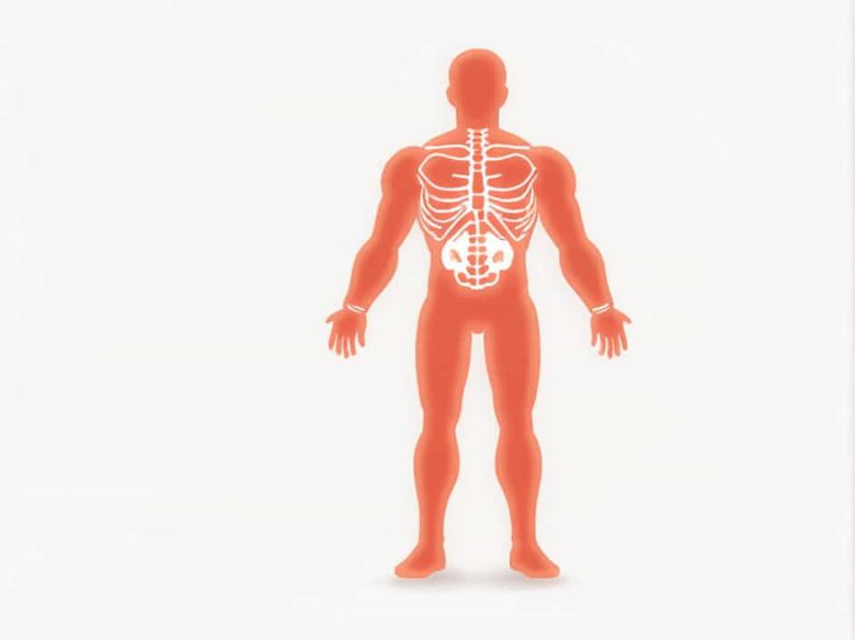
Skeletal muscles are a type of striated muscle tissue attached to bones by tendons. They are responsible for body movement posture and balance. Unlike smooth or cardiac muscles skeletal muscles require conscious control to function.
Key characteristics of skeletal muscles include:
- Voluntary control – They move only when the brain sends a signal.
- Striated structure – They appear striped under a microscope.
- Multinucleated cells – Each muscle fiber contains multiple nuclei.
- Fatigue-prone – They require rest after prolonged activity.
The Role of the Somatic Nervous System (SNS)
How the SNS Controls Skeletal Muscles
The somatic nervous system is part of the peripheral nervous system (PNS) and is responsible for controlling voluntary movements. It sends motor signals from the central nervous system (CNS) to skeletal muscles via motor neurons.
The SNS works in three main steps:
- Sensory Input – The brain receives information about body position and surroundings.
- Decision Making – The brain processes this information and decides on a movement.
- Motor Output – The brain sends a signal through motor neurons to activate the appropriate skeletal muscles.
Components of the Somatic Nervous System
- Motor neurons – Transmit signals from the brain to skeletal muscles.
- Neuromuscular junctions – Sites where motor neurons communicate with muscles.
- Acetylcholine (ACh) – A neurotransmitter that triggers muscle contraction.
Brain Regions Responsible for Voluntary Muscle Control
1. Motor Cortex: The Command Center
The primary motor cortex located in the frontal lobe is the main area responsible for initiating voluntary movements.
- The precentral gyrus of the motor cortex sends signals to motor neurons.
- The motor homunculus represents different body parts with larger areas for hands and face due to their precise movements.
2. Basal Ganglia: Movement Coordination
The basal ganglia a group of deep brain structures help fine-tune movements by preventing unwanted muscle activity. Disorders of the basal ganglia such as Parkinson’s disease lead to tremors and difficulty moving.
3. Cerebellum: Balance and Precision
The cerebellum ensures smooth coordinated movements by adjusting muscle activity. It receives sensory feedback and modifies motor commands accordingly.
4. Brainstem and Spinal Cord: Signal Transmission
The brainstem and spinal cord relay movement instructions from the brain to the body. The spinal cord contains reflex circuits that allow quick reactions without conscious thought.
Motor Neurons and Neuromuscular Junctions
Types of Motor Neurons
Motor neurons transmit movement commands from the CNS to skeletal muscles. There are two types:
- Upper motor neurons (UMNs) – Located in the brain and spinal cord they initiate movement.
- Lower motor neurons (LMNs) – Directly connect to muscles and trigger contractions.
Damage to UMNs causes spastic paralysis while damage to LMNs leads to flaccid paralysis.
Neuromuscular Junctions: Where Nerves Meet Muscles
At the neuromuscular junction motor neurons release acetylcholine (ACh) which binds to muscle receptors and triggers contraction. If ACh release is blocked (as in botulism) muscles become paralyzed.
The Process of Voluntary Muscle Contraction
- Brain Initiates Movement – The motor cortex sends signals to motor neurons.
- Signal Travels via the Spinal Cord – Nerve impulses pass through the spinal cord.
- Motor Neurons Activate Muscles – ACh is released at the neuromuscular junction.
- Muscle Fibers Contract – Muscle filaments (actin and myosin) slide past each other producing movement.
Disorders Affecting Voluntary Muscle Control
1. Neuromuscular Diseases
- Myasthenia Gravis – An autoimmune disorder where the body attacks ACh receptors causing muscle weakness.
- Amyotrophic Lateral Sclerosis (ALS) – A progressive disease that damages motor neurons.
2. Spinal Cord Injuries
Damage to the spinal cord can interrupt signals between the brain and muscles leading to paralysis.
3. Stroke
A stroke can damage the motor cortex affecting voluntary muscle control.
Voluntary control of skeletal muscles is provided by the somatic nervous system which sends signals from the brain and spinal cord to motor neurons. Key brain regions involved include the motor cortex basal ganglia cerebellum and brainstem.
Motor neurons and neuromuscular junctions ensure that movement signals reach skeletal muscles allowing us to perform daily activities. Understanding how voluntary muscle control works can help in diagnosing and treating neuromuscular disorders that impair movement.